
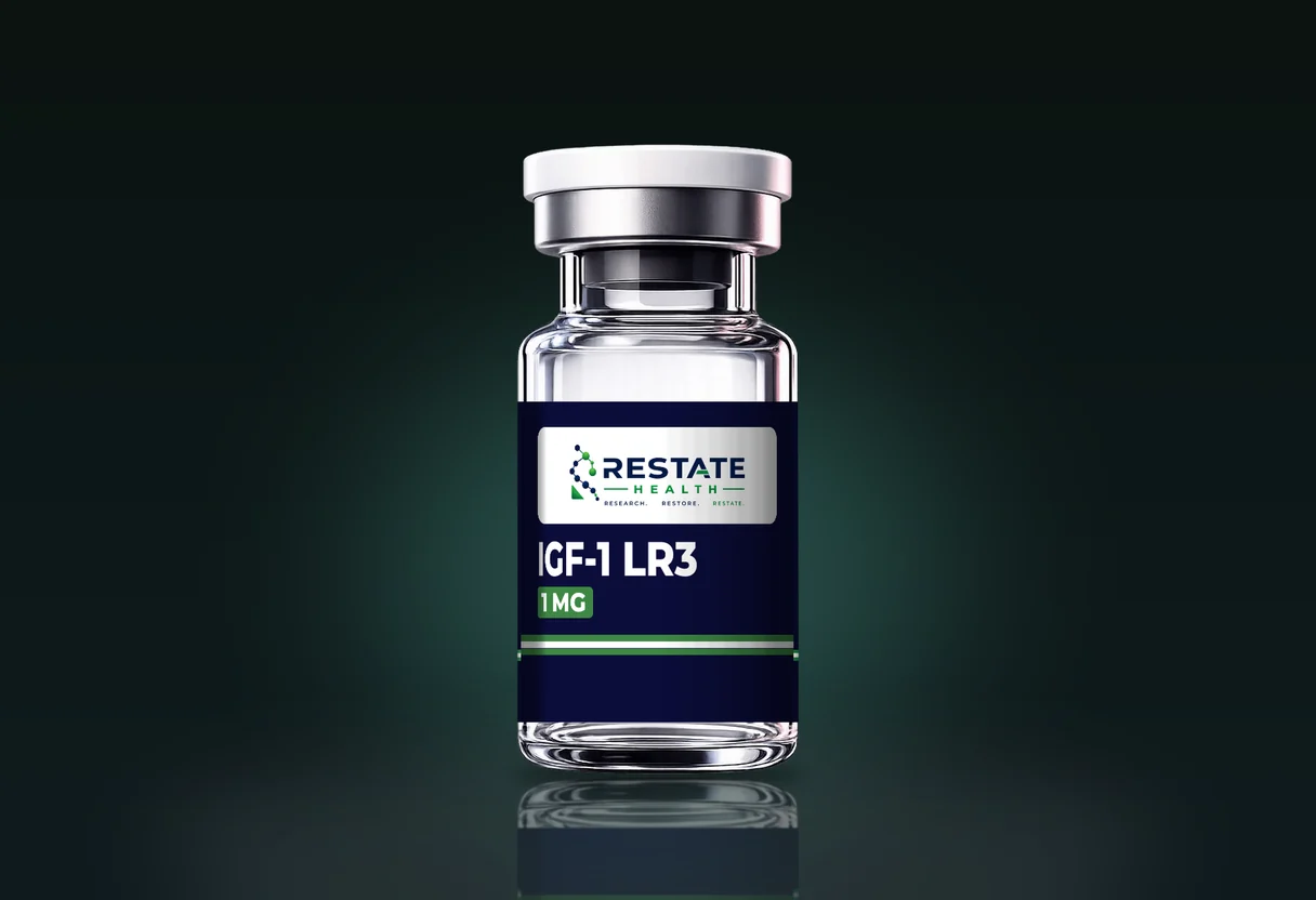
IGF-1 LR3 is the longest single chain in this catalogue: 83 residues, a molecular weight of 9117.60 g/mol, supplied at 1 mg. Those three figures together make it a different kind of purchase from a synthetic peptide. It is a recombinant protein, expressed in Escherichia coli, folded, and held in shape by three disulfide bonds, and the documentation that describes it properly is the documentation of a biologic rather than of a synthesised chain.
This guide covers what the name means residue by residue, why the molecule is made by expression rather than by synthesis, what that changes on the certificate, why endotoxin is a relevant line for an E. coli product, and how a single milligram of carrier-free protein behaves in a vial. Peptide vs protein covers where the line between the two words falls; IGF-1 LR3 sits on the protein side of it.
| IGF-1 LR3 | Catalog specification |
|---|---|
| Vial strengths | 1 mg |
| Sequence | 83-residue recombinant IGF-1 analog with Arg substituted for Glu at position 3 and a 13-residue N-terminal extension; modifications reduce IGFBP affinity and extend circulating half-life relative to native IGF-1 (full sequence per COA). |
| Molecular formula | C400H625N111O115S9 |
| Molecular weight | 9117.60 g/mol |
| Amino acid count | 83 |
| CAS number | 946870-92-4 |
From the Restate Health catalog record. Lot-specific values are on the certificate of analysis.
The parent molecule: IGF-1
Insulin-like growth factor I was sequenced by Rinderknecht and Humbel in 1978, who reported a 70-residue single chain with structural homology to proinsulin. The mature chain, recorded in UniProt as residues 49 to 118 of the P05019 precursor, reads Gly-Pro-Glu-Thr-Leu-Cys-Gly-Ala-Glu-Leu-Val-Asp-Ala-Leu-Gln-Phe-Val-Cys-Gly-Asp-Arg-Gly-Phe-Tyr-Phe-Asn-Lys-Pro-Thr-Gly-Tyr-Gly-Ser-Ser-Ser-Arg-Arg-Ala-Pro-Gln-Thr-Gly-Ile-Val-Asp-Glu-Cys-Cys-Phe-Arg-Ser-Cys-Asp-Leu-Arg-Arg-Leu-Glu-Met-Tyr-Cys-Ala-Pro-Leu-Lys-Pro-Ala-Lys-Ser-Ala.
Two features of that sequence matter for what follows. The third residue is glutamate. And there are six cysteines, at positions 6, 18, 47, 48, 52 and 61, which pair into three disulfide bonds, 6 to 48, 18 to 61 and 47 to 52, in the insulin-like fold. A synthetic chain of this length with six cysteines would have to be folded and oxidised to the right pairing after synthesis, which is one of the two reasons it is not made that way.
What "Long R3" means
Both halves of the name come from the Adelaide group that built the molecule in the early 1990s. King and colleagues, Journal of Molecular Endocrinology 1992, describe expressing IGF-I in E. coli as a fusion protein: the first 46 residues of methionyl porcine growth hormone, then the dipeptide Val-Asn, then IGF-I. The Asn-Gly junction is cleavable with hydroxylamine, so the fusion partner could be cut away, the reduced protein refolded and the product purified. The same system produced two variants with glutamate 3 replaced by glycine or arginine. Both bound very poorly to bovine IGF-binding protein-2 and slightly less well than IGF-I to the type-1 receptor on rat L6 myoblasts, and both were more potent than IGF-I in the cell assays used.
Francis and colleagues, in the same journal later in 1992, took the shorter route that gives the catalogue molecule. They left a fusion partner attached: the first eleven residues of methionyl porcine growth hormone plus Val-Asn, thirteen residues in all, ahead of an IGF-I sequence carrying arginine at position 3. That is Long [Arg3]-IGF-I, written LR3. The paper reports that the hydrophobic N-terminal extension helped the reduced protein fold correctly, and that in L6 rat myoblasts the Long analogues were more potent than authentic IGF-I. Seventy residues plus thirteen gives the 83 in the catalogue record.
The catalogue describes the molecule as an 83-residue recombinant IGF-1 analog with Arg substituted for Glu at position 3 and a 13-residue N-terminal extension, and records that the modifications reduce IGFBP affinity and extend circulating half-life relative to native IGF-1. The binding-protein claim is the one the 1992 papers measure directly. Conlon and colleagues, Journal of Endocrinology 1995, infused LR3 IGF-I into guinea pigs for seven days and reported that plasma IGF-I, IGF-II and IGF-binding protein concentrations fell, that the fractional weights of adrenals, gut, kidneys and spleen increased, and that overall growth was not stimulated. That is the model, and the readouts, behind the sentence in the record.
| IGF-1 | IGF-1 LR3 | |
|---|---|---|
| Residues | 70 | 83 |
| Position 3 | Glu | Arg |
| N-terminal extension | None | 13 residues: Met-pGH(1-11)-Val-Asn |
| Cysteines and disulfides | 6 cysteines, 3 bonds | 6 cysteines, 3 bonds |
| Molecular formula | Not a catalogue item | C400H625N111O115S9 |
| Molecular weight | Not a catalogue item | 9117.60 g/mol |
| Binding to IGF-binding proteins | Binds | Much reduced (King 1992; Conlon 1995) |
| How it is made | Native, or recombinant | Recombinant, E. coli |
IGF-1 from UniProt P05019 and Rinderknecht and Humbel (1978); IGF-1 LR3 from the catalogue record and Francis and colleagues (1992). IGF-1 itself is not a catalogue item.
Why it is expressed rather than synthesised
Solid-phase peptide synthesis builds a chain one residue at a time, and the yield of full-length product falls with every coupling. At 83 residues the crude product is dominated by deletion sequences, and the six cysteines would then have to be oxidised to the correct three pairings out of the fifteen possible. Expression in E. coli sidesteps the first problem entirely, since the cell makes the full chain from a gene, and the 1992 papers show the second problem being managed by refolding the reduced protein, with the N-terminal extension helping.
The consequence for a buyer is that the impurities are different in kind. A synthetic peptide carries deletion and truncation sequences, protecting-group remnants and trifluoroacetate. A recombinant protein carries host-cell proteins and DNA, endotoxin from the E. coli cell wall, misfolded or disulfide-scrambled isomers, and oxidation at the single methionine (position 59 of the IGF-1 chain). The certificate has to be read for the second list, not the first.
What changes on the certificate
| Line | Synthetic peptide | IGF-1 LR3 |
|---|---|---|
| Identity | Intact mass or sequence by fragmentation | Intact mass by electrospray, deconvoluted, near 9117 (average); peptide mapping or N-terminal sequencing. King and colleagues used HPLC and N-terminal sequence analysis |
| Purity | Reversed-phase HPLC area percent | Reversed-phase HPLC and SDS-PAGE; Q6B lists both electrophoretic and chromatographic purity for biotechnological products |
| Folding and disulfides | Not applicable, or one bond | Three bonds; scrambled isomers separate on reversed-phase HPLC; SDS-PAGE run reduced and non-reduced shows aggregates linked by disulfides |
| Process impurities | Trifluoroacetate, residual solvents | Host-cell protein, host-cell DNA, residual process reagents |
| Endotoxin | Relevant where sterility is claimed | Relevant because the source organism is Gram-negative; USP 85 method |
| Content | Net peptide content by amino acid analysis | Protein content, typically by absorbance at 280 nm or amino acid analysis; the molecule has three tyrosines and no tryptophan in the IGF-1 chain |
| Biological activity | Not applicable | Q6B includes a potency test for biotechnological products; a research certificate may or may not carry one |
Certificate lines for a synthetic peptide and for IGF-1 LR3. The test categories for a recombinant protein follow ICH Q6B; the endotoxin method is USP General Chapter 85.
The mass line needs the same care as any large molecule. 9117.60 g/mol is an average mass for the oxidised protein; the fully reduced form would be six hydrogens, about 6 Da, heavier, and at this size the average and monoisotopic values are several daltons apart. A certificate that reports an observed mass should say which basis it is on and whether the sample was reduced, or the comparison is not possible. Molecular weight for buyers covers the two conventions, and they bite harder at 9 kDa than at 1 kDa.
Endotoxin and the E. coli origin
Endotoxin is lipopolysaccharide from the outer membrane of Gram-negative bacteria, and E. coli is one. A protein purified from E. coli starts with endotoxin in the lysate and purification removes most of it; how much remains is a measured quantity, not an assumption. For material that will go into cell culture that figure matters on its own terms, because endotoxin acts on cells independently of the protein under study, which is why Q6B treats it as a process-related impurity to be controlled.
The method is the bacterial endotoxins test of USP General Chapter 85, and endotoxin testing explained covers what it measures and what a result on a certificate does and does not establish. For a synthetic peptide the line is often absent and its absence is unremarkable. For a recombinant protein from E. coli, its absence is a question to ask.
Handling a 1 mg vial of carrier-free protein
One milligram of a 9 kDa protein is about 110 nanomoles. Proteins adsorb to glass and to many plastics, and at low concentration the fraction lost to the surfaces of a vial, a pipette tip and a tube can be a meaningful share of what was bought. The catalogue record does not list a carrier protein, and a carrier-free lyophilisate has nothing in it competing for those surfaces. That is the main practical difference from a 10 mg peptide vial, where the same absolute loss is a rounding error.
The storage line in the record is the catalogue's standard one: lyophilised at -20°C, protected from light and moisture, with the reconstituted solution refrigerated. For a folded protein the freeze-thaw question is sharper than for a short peptide, since repeated freezing promotes aggregation of a molecule that depends on its fold. Freeze-thaw and aliquoting covers dividing a solution once rather than cycling a single vial, and that advice was written with proteins like this one in mind.
What to check before ordering
- An observed intact mass near 9117.6 with the basis stated, and a purity figure from reversed-phase HPLC with SDS-PAGE alongside it.
- Some evidence of correct folding: a single main peak on reversed-phase HPLC, and a non-reduced SDS-PAGE lane without higher bands.
- An endotoxin result, with the method, for a product expressed in E. coli.
- A protein content figure and its method, since the label mass is lyophilised solid.
- Whether a carrier protein is present. The record lists none.
- That the certificate names the lot on the vial.
The material is supplied strictly for laboratory research. What this guide can settle from documentation is narrower than the literature and more useful to a buyer: which molecule is in the vial, whether it is folded, how pure it is, how much of it there is, and whether the lot in hand is the one the certificate describes.