
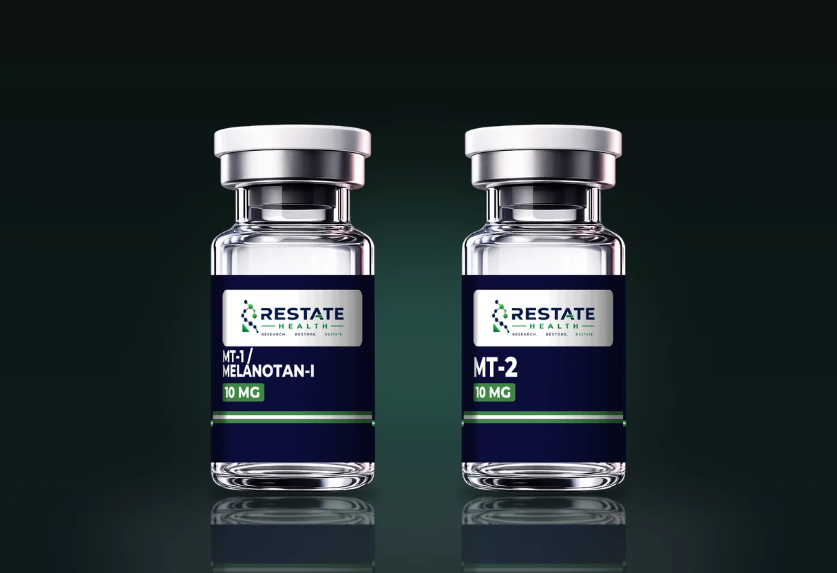
MT-1 and MT-2 sit next to each other in every melanocortin listing and are often spoken of as a pair. Structurally they are not a pair. MT-1, also recorded as afamelanotide and [Nle4-D-Phe7]-alpha-MSH, is a linear 13-residue peptide. MT-2 is a seven-residue fragment closed into a ring by a side-chain lactam. One is the full hormone length with two substitutions; the other is about half of it, bridged.
This guide takes the two apart residue by residue, follows the design history from the 1980 paper to the 1989 paper, and sets out what cyclisation changes for stability and for the mass spectrometry a certificate rests on. It does not cover receptors or what either compound does in any model; melanocortin peptides explained covers the family, the five receptor subtypes and the published binding work, and none of that is repeated here.
| Compound | Vial strengths | Molecular weight | Amino acids |
|---|---|---|---|
| MT-1 / Melanotan-I | 10 mg | 1646.87 g/mol | 13 |
| MT-2 | 10 mg | 1024.18 g/mol | 7 |
From the Restate Health catalog records. Lot-specific values are on each certificate of analysis.
The parent sequence and the 1980 substitution
Alpha-MSH is Ac-Ser-Tyr-Ser-Met-Glu-His-Phe-Arg-Trp-Gly-Lys-Pro-Val-NH2, thirteen residues with an acetylated N-terminus and an amidated C-terminus, as written out in Al-Obeidi and colleagues' 1989 paper. Sawyer and colleagues reported in 1980 that heating the native hormone in alkali prolonged its activity in a frog skin assay, and that gas chromatography of the hydrolysed peptide showed partial racemisation, particularly at positions 4 (methionine) and 7 (phenylalanine).
They then made the substitution deliberately: norleucine at position 4 in place of methionine, and D-phenylalanine at position 7 in place of the L-residue. The resulting [Nle4, D-Phe7]-alpha-MSH was resistant to degradation by serum enzymes and showed prolonged and markedly increased activity in the frog skin bioassay and in mouse melanoma adenylate cyclase and tyrosinase assays. That molecule is MT-1. The catalogue sequence, Ac-Ser-Tyr-Ser-Nle-Glu-His-D-Phe-Arg-Trp-Gly-Lys-Pro-Val-NH2, is the native hormone with exactly those two changes.
From thirteen residues to seven: the 1989 lactam
Al-Obeidi, Castrucci, Hadley and Hruby took the 4-10 fragment of that analogue and closed it into a ring. In their 1989 paper the Glu at position 5 became Asp, the Gly at position 10 became Lys, and a lactam bond was formed between the Asp side-chain carboxyl and the Lys side-chain amine, either in solution or on the solid-phase support. The acetyl at the N-terminus and the amide at the C-terminus were kept. Seven cyclic peptides were made and assayed in frog and lizard skin; the Asp5-Lys10 analogue, with a 23-membered ring, was about 90 times as potent as alpha-MSH in the lizard assay and showed prolonged activity, while the smaller rings of 20 to 22 members did not.
That analogue, Ac-Nle4-cyclo[Asp5, D-Phe7, Lys10]-alpha-MSH-(4-10)-NH2 in the nomenclature Hruby and colleagues used in 1995, is the catalogue sequence for MT-2: Ac-Nle-cyclo[Asp-His-D-Phe-Arg-Trp-Lys]-NH2. So MT-2 is not a trimmed MT-1. It is the central seven residues of MT-1 with two further substitutions made specifically so that a ring could be closed.
Residue by residue
| Position | Alpha-MSH | MT-1 | MT-2 |
|---|---|---|---|
| 1 | Ac-Ser | Ac-Ser | absent |
| 2 | Tyr | Tyr | absent |
| 3 | Ser | Ser | absent |
| 4 | Met | Nle | Ac-Nle (N-terminus) |
| 5 | Glu | Glu | Asp (lactam to Lys10) |
| 6 | His | His | His |
| 7 | Phe | D-Phe | D-Phe |
| 8 | Arg | Arg | Arg |
| 9 | Trp | Trp | Trp |
| 10 | Gly | Gly | Lys-NH2 (lactam to Asp5; C-terminus) |
| 11 | Lys | Lys | absent |
| 12 | Pro | Pro | absent |
| 13 | Val-NH2 | Val-NH2 | absent |
Alpha-MSH as written in Al-Obeidi and colleagues (1989); MT-1 and MT-2 as recorded in the catalogue. Position numbers follow alpha-MSH.
Read down the MT-2 column and the shared core is visible: His-D-Phe-Arg-Trp at positions 6 to 9 is identical in all three. Everything else about MT-2 is scaffolding built so that core holds a particular shape.
Two masses, two formulas
The catalogue records give MT-1 as C78H111N21O19, 1646.87 g/mol, CAS 75921-69-6, and MT-2 as C50H69N15O9, 1024.18 g/mol, CAS 121062-08-6. PubChem agrees: CID 16197727 (afamelanotide) lists 1646.8 with a monoisotopic mass of 1645.84, and CID 92432 (Melanotan II) lists 1024.2 with a monoisotopic mass of 1023.54. Average and monoisotopic values sit about one dalton apart at these sizes, so a certificate should say which basis its observed mass is on before the comparison is made; molecular weight for buyers covers the two conventions.
The 622.69 g/mol between them is the six residues MT-2 does not carry (Ser, Tyr, Ser at one end and Lys, Pro, Val at the other), adjusted for the Glu-to-Asp and Gly-to-Lys changes and for the water lost when the lactam closed. That last item is the one a certificate can catch: open-chain MT-2 that never cyclised is one water heavier than the ring, which the melanocortin guide works through.
What cyclisation does to stability
A linear peptide can take up many conformations in solution; a ring can take up far fewer. That constraint is the design purpose, and it is also why cyclisation is synthetically demanding. White and Yudin's 2011 review of peptide macrocyclisation puts the difficulty plainly: the activated chain must adopt an entropically disfavoured pre-cyclisation conformation before the ring can form, which is why incomplete cyclisation is a realistic impurity in any cyclic product and why the plus-18 check matters.
The ring also changes what a single backbone cleavage does. Cut a linear peptide once and it falls into two pieces of lower mass. Cut the bridged segment of MT-2 once and the molecule opens but does not divide: it is now a linear peptide of the same formula plus one water, still one piece. The Al-Obeidi paper's observation that the 23- and 24-membered rings showed prolonged residual activity in both skin bioassays while the smaller rings did not is the experimental face of that structural difference, measured in amphibian and reptile skin preparations.
MT-1 has its own stabilisation, which is the 1980 one: a D-residue at the position where the native hormone was most readily racemised, plus norleucine in place of an oxidisable methionine. Sawyer and colleagues reported the resulting analogue as resistant to serum enzymes. So both molecules are stabilised; they are stabilised by different means, and the means leave different marks on a certificate.
What cyclisation does to the fragment ladder
Identity by mass spectrometry comes in two strengths: an intact mass, which shows the composition is consistent with the molecule, and a fragment spectrum, which reads the sequence. Mass spectrometry and peptide identity sets out the difference. For MT-1 the second is straightforward: a 13-residue linear chain fragments into b- and y-type series that step along the sequence one residue at a time.
For MT-2 it is not. Ngoka and Gross showed in 1999 that collisionally activating a protonated cyclic peptide opens the ring at random positions first, so the spectrum is a superposition of fragment series from several different linear openings, all giving acylium ions of the same m/z and difficult to assign. Their solution was multistage tandem MS in an ion trap, selecting one ring-opened ion and stripping residues from it stage by stage. The practical consequence for a buyer: a certificate that claims sequence confirmation by MS/MS for a cyclic peptide should say how the ring was handled, and a certificate that offers only an intact mass for MT-2 has shown composition, not sequence.
There is a second limit that applies to both. Norleucine, leucine and isoleucine share the formula C6H13NO2 and therefore the same residue mass, so neither an intact mass nor an ordinary fragment ladder distinguishes Nle at position 4 from Leu. That is a plain consequence of the elements involved, and it means the norleucine in both catalogue sequences is established by the synthesis record rather than by the spectrometer.
D-Phe: the residue mass spectrometry cannot see
D-phenylalanine and L-phenylalanine have the same formula, the same mass and the same fragment masses. A mass spectrometer, used in the ordinary way, is blind to the difference; Tao and Cooks' 2003 review of chiral analysis by MS exists precisely because special approaches are needed to see it at all. So the D-Phe at position 7, which both catalogue sequences specify and which the 1980 paper identified as the stabilising change, is not something either certificate's MS line can confirm.
Where it is controlled is in synthesis. The D-residue enters as a protected building block during solid-phase peptide synthesis, and whether the finished peptide carries D or L at position 7 depends on which building block was coupled and on how much racemisation the coupling conditions allowed. Where it can be checked after the fact is the method Sawyer and colleagues used in 1980: hydrolyse the peptide to free amino acids and run a chiral separation, which reports the D-to-L ratio at each residue. A supplier who can produce that result for a melanocortin analogue has answered a question the MS line cannot.
What to check on a certificate for each
- MT-1: intact mass reconciling with 1646.87 (average) or 1645.84 (monoisotopic), basis stated; a fragment ladder if sequence confirmation is claimed; purity by HPLC; and, if offered, a chiral amino acid analysis for position 7.
- MT-2: intact mass reconciling with 1024.18 (average) or 1023.54 (monoisotopic), with nothing near 1042 that would indicate open-chain material; a statement of how MS/MS handled the ring if sequence confirmation is claimed; purity by HPLC; and the same chiral question.
- Both: the lot number on the vial against the certificate, and the salt form by asking, since it is rarely on the document.
Both are supplied as 10 mg lyophilised vials at $64.40, stored at -20 °C and protected from light and moisture, so the buying decision between them is not price or handling. It is which molecule the work calls for, and that is a question for the literature rather than for a catalogue.
In short
MT-1 is the native hormone's thirteen residues with Nle at 4 and D-Phe at 7, the 1980 design. MT-2 is residues 4 to 10 of that analogue with Asp at 5 and Lys at 10 bridged into a 23-membered lactam ring, the 1989 design. The ring constrains shape, survives a single cleavage, and scrambles an ordinary fragment spectrum; the D-Phe both carry is invisible to the spectrometer and lives in the synthesis record. Reconcile the masses on the stated basis, ask how the ring was sequenced, and read the melanocortin guide for everything about receptors.