
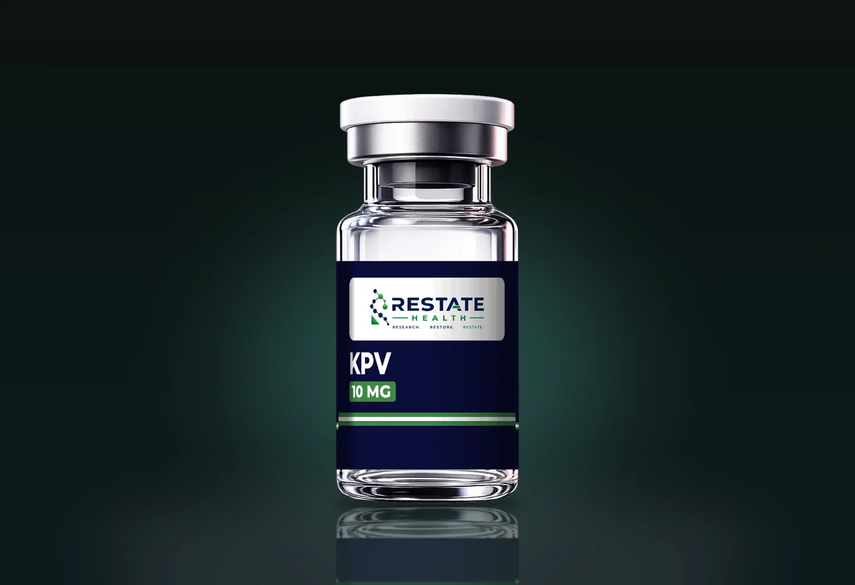
KPV is about as small as a peptide gets while still being called one. It is three residues, lysine, proline and valine, with a molecular weight of 342.43 g/mol in the catalogue record, and it corresponds to the last three residues of the thirteen-residue hormone alpha-melanocyte-stimulating hormone, which is why the record lists α-MSH(11-13) as a synonym. The catalogue supplies it in a single 10 mg strength.
Its size is the whole story for a buyer. A tripeptide sits in the mass range of ordinary small molecules, and that changes what a certificate can prove about it, how much of the solid in a vial is actually peptide, and what the melanocortin literature does and does not say about it. This guide takes those in turn. The material is supplied strictly for laboratory research.
| KPV | Catalog specification |
|---|---|
| Vial strengths | 10 mg |
| Sequence | Lys-Pro-Val |
| Molecular formula | C16H30N4O4 |
| Molecular weight | 342.43 g/mol |
| Amino acid count | 3 |
| CAS number | 67727-97-3 |
From the Restate Health catalog record. Lot-specific values are on the certificate of analysis.
Where the three residues come from
Alpha-MSH is a 13-residue peptide cut from the larger precursor pro-opiomelanocortin. Its C-terminal tripeptide, Lys-Pro-Val, was examined on its own from the late 1980s onward. Hiltz, Catania and Lipton reported in 1991 on analogues of alpha-MSH(11-13) and on how changes to their stereochemistry altered activity in the inflammation assays they used (Peptides 1991), which is also the first reason a buyer should care about chirality for this compound.
Catania, Gatti, Colombo and Lipton's 2004 review of melanocortin receptors as a route to controlling inflammation sets the tripeptide in the context of the whole melanocortin family (Pharmacol Rev 2004). Melanocortin peptides explained covers that family as a catalogue class.
One point from that literature bears on how KPV is described. The 2008 cell work discussed below proposes a transporter-mediated route into cells rather than action at a melanocortin receptor. KPV is a fragment of alpha-MSH by sequence; it is not a small version of it by mechanism, and the two should not be conflated on a product page or in a record.
What the published models describe
Two 2008 papers are the ones most often cited for KPV, and both name their systems.
- Dalmasso and colleagues worked in human intestinal epithelial cell lines, Caco2-BBE and HT29-Cl.19A, and in Jurkat T cells, where they report that KPV at nanomolar concentrations reduced activation of NF-kappaB and MAP kinase signalling after cytokine stimulation, and that uptake ran through the di/tripeptide transporter PepT1. They then examined two mouse colitis models, induced with DSS and with TNBS, with KPV added to the drinking water (Gastroenterology 2008).
- Kannengiesser and colleagues examined the tripeptide in murine models of inflammatory bowel disease the same year (Inflamm Bowel Dis 2008).
That is what the literature says and where. The catalogue about line summarises it as anti-inflammatory signalling studied in gut and mucosal inflammation research models. Nothing in it is a certificate claim, and nothing on a certificate speaks to it.
Why 342 Da is weak evidence of identity on its own
For a 4,000 Da peptide, a mass that reconciles with the expected figure to within a dalton is strong evidence that the right molecule is present, because once a sequence is specified the number of plausible peptides near that mass is small. At 342 Da the situation is different. Kind and Fiehn's 2007 analysis of molecular-formula assignment from accurate mass shows how many candidate formulas fit a measured mass even at high accuracy and how the count grows with mass tolerance (BMC Bioinformatics 2007). In the small-molecule range those candidates include a great many compounds that are not peptides at all.
There is a sharper problem that needs no formula search. The six possible orderings of lysine, proline and valine (KPV, KVP, PKV, PVK, VKP and VPK) all have the formula C16H30N4O4 and all weigh 342.43 g/mol. An intact mass cannot tell them apart. Nor can it distinguish the all-L peptide from one carrying a D-residue, since stereoisomers share a mass, and the 1991 paper's subject was precisely such stereoisomers.
What separates them is fragmentation. Tandem mass spectrometry breaks the peptide along its backbone and reads the pieces; the b- and y-ion masses differ between KPV and, say, VPK because the residues sit in a different order. For a tripeptide the fragment set is small and easy to interpret, which makes the method cheap to run and decisive. Mass spectrometry and peptide identity covers the difference between an intact mass and a fragment ladder in general.
| Method | What it reports for KPV | What it rules out | What it cannot rule out |
|---|---|---|---|
| Intact mass | An ion consistent with 342.43 (average) or 342.23 (monoisotopic) | A wrong formula; gross contamination | The five other K/P/V orderings; D-residue isomers; any non-peptide with the formula C16H30N4O4 |
| Tandem MS (MS/MS) | Backbone fragments whose masses fix the order Lys-Pro-Val | Sequence permutations; most isobaric non-peptides | D- versus L-residues |
| Chiral amino acid analysis | The L or D configuration of each residue after hydrolysis | D-residue isomers | Residue order |
| Retention time against a reference standard | Co-elution with a known KPV standard | Most isomers, if the method separates them | Anything the method does not resolve |
What each identity method can and cannot establish for a 342 Da tripeptide. The average mass is the catalogue record; the monoisotopic mass is PubChem CID 125672.
A certificate that gives only an intact mass for KPV has shown that something with the right formula is in the vial. One that adds MS/MS, or co-elution with a reference standard, has shown that it is KPV.
Purity on a molecule this small
HPLC purity is a share of detector response, and UV detection of peptides at 214 to 220 nm reads the amide bonds. KPV has two of them and no aromatic residue, so it absorbs weakly and gives a modest signal. Impurities carrying aromatic groups would be over-represented in the trace, and non-absorbing contaminants, salts and water are invisible to it. The catalogue lists ≥99% by HPLC, which is a sound figure as far as it goes; what it does not go to is the share of the solid that is peptide.
Net content and the counterion share
This is where a tripeptide differs most from a long peptide. The counterion attaches to basic sites, and KPV has two: the lysine side chain and the free N-terminal amine. Two trifluoroacetates weigh 228 Da. On a 4,400 Da peptide two counterions are a rounding error. On a 342 Da peptide they are two-fifths of the salt.
| Salt form | Counterion mass added | Mass of the salt (g/mol) | Peptide share by mass | Peptide in 10 mg of dry salt |
|---|---|---|---|---|
| Free base, no counterion | 0 | 342.43 | 100% | 10.0 mg |
| Mono-acetate | 60.05 | 402.48 | 85% | 8.5 mg |
| Di-acetate | 120.10 | 462.53 | 74% | 7.4 mg |
| Mono-trifluoroacetate | 114.02 | 456.45 | 75% | 7.5 mg |
| Di-trifluoroacetate | 228.04 | 570.47 | 60% | 6.0 mg |
Arithmetic from the catalogue molecular weight of 342.43 g/mol, using 114.02 g/mol for trifluoroacetic acid and 60.05 g/mol for acetic acid. The salt form actually supplied is on the certificate or available on request; these rows are illustrations, not catalogue records.
The last column assumes the vial holds 10 mg of salt with no water. Water widens the gap. A certificate that states net peptide content answers the question directly; one that states only a label mass and a purity percentage leaves an uncertainty of up to 40% in how much KPV is present. Net peptide content covers how the figure is measured, and TFA and acetate salt forms covers why the counterion matters beyond mass, particularly in cell work, where the trifluoroacetate a small peptide carries with it arrives at a proportionally high concentration.
Handling, from the catalogue record
The record lists KPV as a lyophilised powder, stored at -20°C and protected from light and moisture, reconstituted in bacteriostatic water with the solution refrigerated at 2-8°C, and soluble in bacteriostatic water with the COA to be consulted for specific solvent recommendations. A charged tripeptide with no aromatic, cysteine, methionine or asparagine residue is about as soluble and as stable as peptides come, and the proline protects the Lys-Pro bond from many proteases. The handling risks here are small compared with the documentary ones above.
What to ask for on a KPV certificate
- An identity method beyond intact mass: MS/MS fragments, or co-elution with a reference standard.
- The mass basis, average or monoisotopic, since 342.43 and 342.23 are both correct and differ by 0.2 Da.
- The salt form, and ideally a net peptide content figure, because the counterion share can approach 40%.
- The lot number matching the vial, as always.
Small molecules are easy to make and easy to mis-describe. The paperwork that settles which is the case is cheap for a supplier to produce, and a buyer is entitled to ask for it.